
A promising biomarker in neurodegenerative disease and traumatic brain injury is the neurofilament light chain protein (NfL), which provides a sensitive measurement of neuroaxonal damage, regardless of cause. Clinical utility includes Tramatic Brain Injury
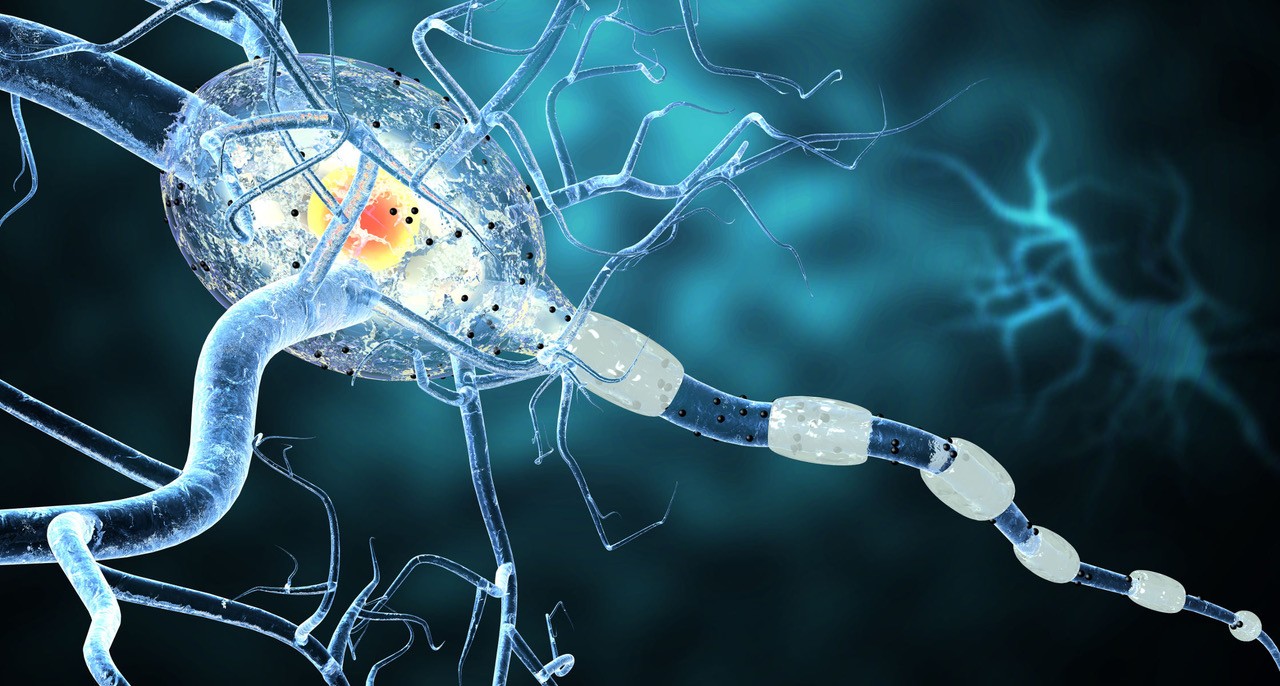
Neurofilaments are the main cytoskeletal structure proteins in neurons. They are about 10 nm in diameter, thicker than actin and thinner than myosin, and are thus classed as intermediate filaments (IFs). Neurofilaments comprise four different subunits, the stoichiometry of which varies depending on the maturity of the neuron. Three of these subunits, NfL (light), NfM (middle) and NfH (heavy), are type IV IF proteins and are present always, and the fourth is either α-internexin (also type IV) or peripherin (type III), for the central and peripheral nervous systems, respectively (see Figure 1).

Figure 1. Subunits of neurofilaments.
Axonal injury and neurodegeneration lead to the appearance of neuronal proteins in the cerebrospinal fluid (CSF). NFs, being abundant proteins exclusively expressed in neurons, may serve as a marker of neuronal degradation. Numerous studies indicate that NFs can be markers of acute conditions (stroke or trauma), as well as a variety of neurological diseases. Monitoring NfL levels in CSF or blood is useful in clinical diagnostic evaluation for predicting the progression of various acute and chronic neurological diseases, as well as for assessing treatment efficacy.
Advanced ImmunoChemical offers several monoclonal antibodies (MAbs) for the development of a highly sensitive and specific NfL immunoassay. MAbs recognize NfL in CSF with high specificity and sensitivity. Pairs of MAbs (Table 1) effectively recognize both recombinant and endogenous NfL and may be used for a variety of immunoassays, such as direct EIA, indirect EIA, and sandwich-type immunoassays with a level of detection at10 pg/ml (Figure 2) and with good correlation to a commercially available reference assay.
Table 1. Pair recommendations for Anti-Neurofilament Light antibodies.


Figure 2. Calibration curve for the CLIA assay prototype NF79-NF71.
The identification and quantification of axonal damage could allow for the improvement of diagnostic accuracy and prognostic assessment. New immunoassays able to detect biomarkers at ultralow levels have allowed for the measurement of NfL in blood, thus making it possible to easily and repeatedly measure NfL for monitoring diseases’ courses. Evidence that both CSF and blood NfL may serve as diagnostic, prognostic and monitoring biomarkers in neurological diseases is progressively increasing, and NfL is one of the most promising biomarkers to be used in clinical and research settings.
For more details: click below